Evolution and development of brainstem sound localisation circuits
Our view of the world, how we move around, how we process the sound information that comes in through our ears or the visual information we detect with our eyes, is all done by circuits of neurons in the brain. These are groups of specific types of neurons, that are connected to each other in specific ways and that receive and send specific inputs and outputs. How are these circuits built during development? How have they evolved? How are the different neuronal types born and become their adult selves?, how do they migrate to their final positions? and how do they extend projections to build the circuit’s connections? These are all crucial questions in neuroscience. Knowing how a circuit is built teaches us a lot about its function, and how and why it may not be working properly.
We are addressing these questions by studying the development and evolution of sound localisation circuits in the brainstem. We are using fate mapping techniques in developing chicken embryos, to trace the different neuronal lineages and combining this with gene expression profiling to better follow their development. We are then comparing the differences and similarities of how sound localisation circuits develop in birds (chicken) and mammals (mouse) to better understand how circuits that perform similar functions have evolved independently.
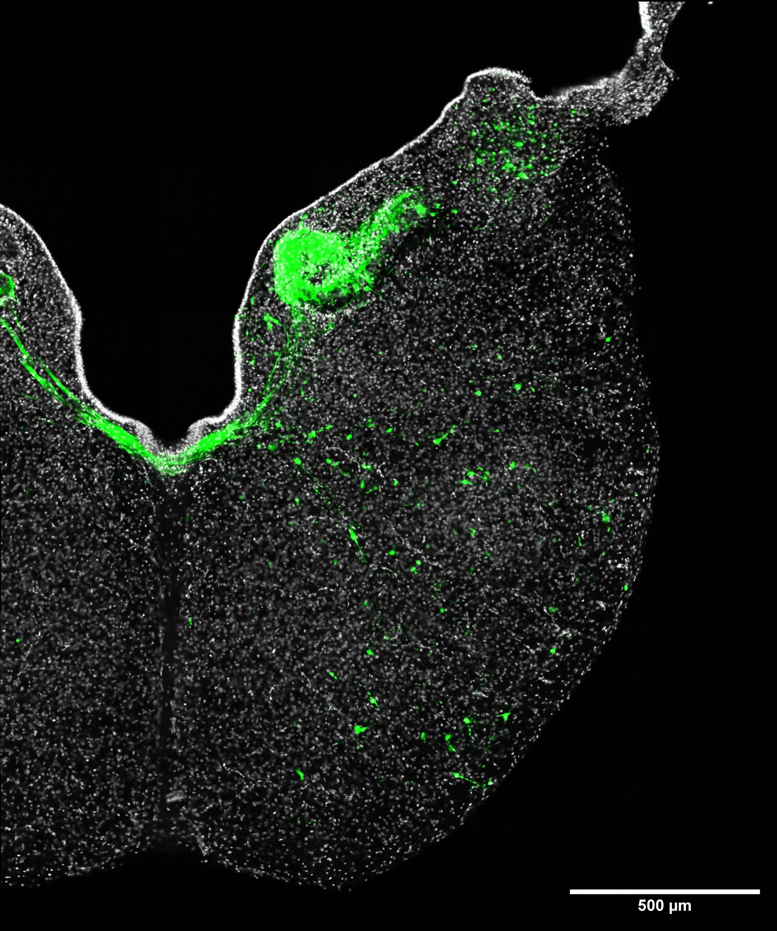
One side of chicken embryo hindbrain transverse section. The sample was collected from chicken embryo at embryonic day 10. The hindbrain is an essential structure in the developing embryo which give rise to the cerebellum and the brainstem in chicken. Neurons that are developed from progenitor cells which express transcription factor, Atoh1, are labelled by green fluorescent protein through electroporation, and are shown in green color here. Cell nuclei are counterstained with DAPI and are shown in gray color.